There is a natural curiosity in engineers to understand exactly how things work.
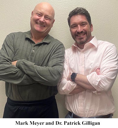 Mark Meyer, 63, an Albuquerque resident who works at Sandia National Laboratories as a field engineer, was very curious about the workings of the hip joint and to understand why his right hip was causing so much pain.
This was not Meyer’s first problem involving his hips. In 2010, he underwent surgery to replace his left hip, but was not at all pleased with the results and recovery.
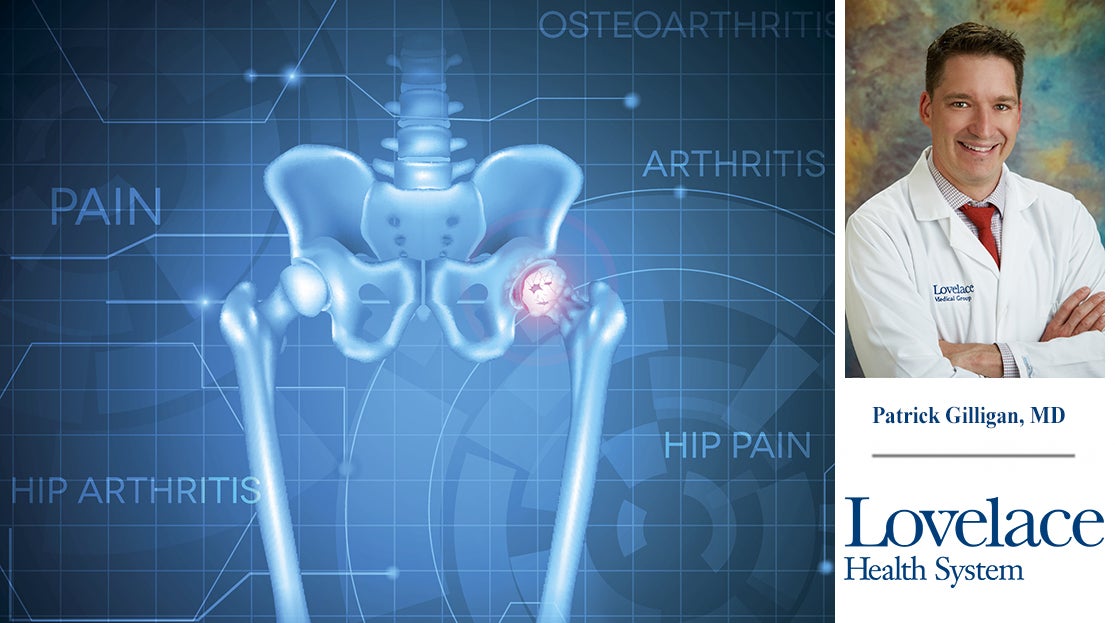
In 2023, when he found walking normally both difficult and very painful, he turned to Patrick Gilligan, M.D., an orthopedic surgeon with Lovelace Medical Group.
After X-rays were taken during an office visit, Dr. Gilligan’s diagnosis was that Meyer’s right hip joint had osteoarthritis.
Osteoarthritis is a degenerative joint disease commonly called “bone-on-bone” or “wear and tear” arthritis. When the cartilage between the bones wears down and disappears after decades of rubbing together, the bones start to break apart.
The hip joint, one of the largest joints in the body, is a “ball and socket” joint. The upper end of the femur (thigh bone) meets the pelvis to create the joint. The “ball” at the end of the femur is called the femoral head and fits into the “socket” (the acetabulum) of the pelvis.
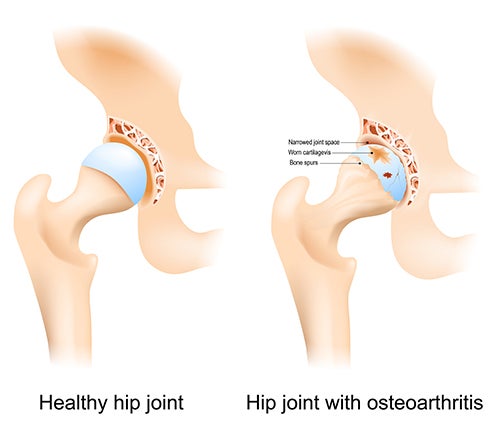 In between the bones is a layer of cartilage which acts as a slippery surface that allows the ball to glide and rotate smoothly when the leg moves. The labrum, a strong cartilage that lines the outer rim of the socket, provides stability.
Dr. Gilligan talked at length with Meyer about the procedure, such as how to prepare, what was going to happen and what to expect afterward – details that the engineer appreciated. “Dr. Gilligan was very thorough in his explanation,” Meyer said.
Hip replacement surgery
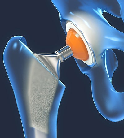 After making an incision over the thigh, Dr. Gilligan removed damaged sections of the hip joint: the femoral head (ball) at the top of the femur (thigh bone) and the acetabulum (socket). He then replaced these damaged sections with new parts constructed of metal, ceramic and very hard plastic. The smoothness of these new parts will help reduce pain and improve Meyer’s mobility.
“My surgery and recovery could not have gone any better,” said Meyer.
Soon thereafter, Meyer began his rehabilitation program to stretch and strengthen his right leg. A key element of his rehabilitation program was to strengthen the gluteal muscles that make up the buttock area.
“Meyer underwent a strong walking program,” recalled Dr. Gilligan. “This activity strengthened his glutes, which protects the hip, keeps it stable, assists with balance, and helps the joint last as long as possible.”
“The physical therapy sessions I attended at Lovelace Medical Center were the best I have ever participated in,” said Meyer enthusiastically. “I am already better than just back to normal. I’m driving now and returned to work after only three weeks.”
“Thank you, Dr. Gilligan and team!”
If you are experiencing pain in your knees, hips or other areas, call Lovelace Medical Group Orthopedics at 505.727.4430. You can also read more and schedule an appointment on our Orthopedic Services web page.